
UCL Microstructure Imaging Group
We use mathematical and computational modeling together with cutting-edge machine learning and parameter estimation techniques to engineer new biomedical imaging techniques. Our primary focus is on magnetic resonance imaging (MRI) for medicine. Specifically, our techniques enhance understanding and early detection of neurological conditions, such as Alzheimers diseases, other dementias, multiple sclerosis, and psychiatric disorders, and assist in the diagnosis and treatment assignment for solid cancers. The microstructure imaging paradigm uses model-based approaches to estimate tissue features traditionally accessible only through invasive biopsy and histology. The ultimate aim is replace such procedures, which currently underpin the gold-standard diagnosis of most cancers and many neurological disorders, with non-invasive imaging. Our Research page details our general approach and specific work.
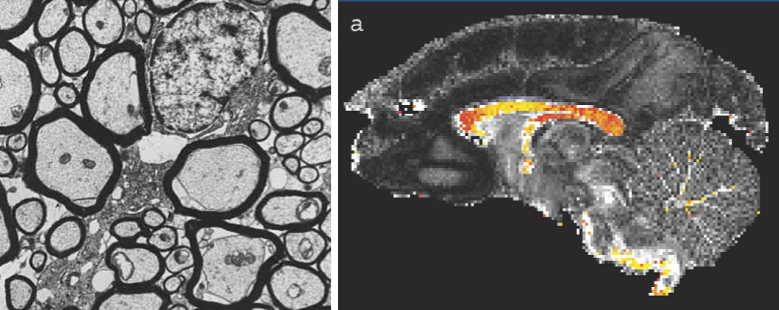 ActiveAx is an example of microstructure imaging in the brain. The technique estimates histological features of white matter tissue (the brains communication network), such as the size, configuration, and density of nerve fibres. The image on the left shows a histological image of white matter from electron microscopy, each axon fibre is a few micrometers in diameter. The image on the right is a map of average axon diameter in each pixel (approximately one millimeter squared) obtained non-invasively from MRI.
Newsletter
Microstructure Imaging Group | Rss / RssFeed
- Rss / Nature Reviews Neurology Research Highlights feature MIG work (Grussu F et al, ACTN 2017)
- Rss / Neurite dispersion: a new marker of multiple sclerosis spinal cord pathology? - Grussu F et al, Ann Clin Transl Neur 2017
- Rss / Image quality transfer and applications in diffusion MRI - Alexander et al. Neuroimage (2017) 152:283-298
- Rss / Apparatus for histological validation of MRI in prostate - Bourne et al., 2017
- Rss / Danny Alexander and Andrada Ianus awarded ISMRM fellowships.
- Rss / Machine learning based compartment models with permeability - Nedjati-Gilani et al.